These images are the abdomen of a 35 year old male. He came in with abdomen pain and had an abnormal CT scan and ultrasound. The patient has a mass in his liver. It is large measuring 7x6cm. The radiologist read this hepatic lesion to most likely be a giant hemangioma due to its enhancement characteristics. In the images I provided you can clearly see the pathology in the patients liver. I found this exam to be very interesting because I have not seen a lot of abdomen scans in MRI. CT is usually the first choice for an abdomen scan because MRIs can be very time consuming and the pt has to do numerous breath holds and may not be in a suitible condition to do so especially if they are in a lot of pain.
Sunday, December 13, 2009
MRI ABDOMEN W/WO
These images are the abdomen of a 35 year old male. He came in with abdomen pain and had an abnormal CT scan and ultrasound. The patient has a mass in his liver. It is large measuring 7x6cm. The radiologist read this hepatic lesion to most likely be a giant hemangioma due to its enhancement characteristics. In the images I provided you can clearly see the pathology in the patients liver. I found this exam to be very interesting because I have not seen a lot of abdomen scans in MRI. CT is usually the first choice for an abdomen scan because MRIs can be very time consuming and the pt has to do numerous breath holds and may not be in a suitible condition to do so especially if they are in a lot of pain.
Wednesday, December 9, 2009
CONTRAST ENHANCEMENT
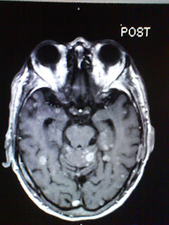
This is an exam I did earlier in the semester that I found interesting. The patient was an 85 yr old man with history of lung cancer. The patient had a screening MRI of the brain about six months prior to the one we did in October to observe for mets due to mental status changes. His GFR was low and the radiologists had the tech do the study without contrast when usually anyone with a cancer history has studies with unless their are contradictions like kidney function. This time the patients GFR was still low but not as low as it was and this radiologist gave us the go ahead to inject contrast due to the patients history. These images are not the best because I took them off the screen at work with my camera phone but you can still see what I am talking about. In the pre images you can see maybe one are that might be something but might be atrophy due to age, it is uncertain if it is a pathology or metastatic disease. Then the injection is given and all I could say was oh my gosh!! All I could keep comparing this man's brain to was a dalmation dogs spots. It was unreal what you could see with the contrast enhancement that you couldn't without it! I wondered if all the mets had been there six months prior when contrast was not used? Look at the images and you will see why I was so shocked at the differences. They didn't upload in the order I wanted them to but the bottom two are the pre images.
Monday, December 7, 2009
MRI
Clinicals are still going smoothly. I am learning a lot. We have two new radiologist and one of them has a thing for MRI so he has been changing many of our protocols. We have also done a lot more abdomen MRIs than normal so I am beginning to get very familiar and comfortable with them. We are using a new contrast called Eovist that is specially designed for liver lesion enhancment. The magnet we work on is a Philips Acheiva 1.5T. It is a closed magnet but seems more open than others because it has a short bore. The short bore is very nice for the patients but sometimes it causes fat sat issues with our images. The lead tech said they have had problems with it ever since they got it and Philips said it is just something the short bore magnets run into sometimes. It usually only comes up when scanning feet and breast. One end of the foot might fat sat and the other not. It is really frustrating when doing a breast MRI because they are so lengthy. The majority of the time the fat sat does not work properly on patients with breast implants and we have to make different adjustments with techniques and factors and just keep running scans and keep changing factors until it works. Not the same changes work with every patient, it is like certain things work according to the patients tissue make up. I'm beginning to learn what adjustments to make with the shim box, spir and spair, etc. All in all it is a great learning experience having to know what to do when something doesn't go completely as planned.
Thursday, November 12, 2009
CERVICAL MRI W/WO
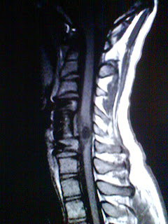
This patient's history really got to me and it is a very interesting exam to see. The images I posted do not look like the original because I took these with my camera phone off of the computer screen so the T1 and T2 weighted images are not windowed properly but it was the best I could get. You can still clearly see the pathology I am talking about. This young man is only 22 years old, the same age as my brother, so that is why I think I put so much emotion into this study. The parents of this patient were practically having to do everything for him. He had a motorized wheelchair with a joy stick because he had very little use of his upper extremities and had been paralized from the waist down. I assumed he was born like this until I screened him and got his history. Last summer he (being over six feet tall himself) dove head first into an above ground swimming pool and suffered severe trauma to his cervical spine. He had to have surgery fusing C4 to C6 with a large strut graft and fusion plate. His injury paralized him. I felt so much sorrow for him and his family because he was an average everyday young man and because of his injury he will have to endure many hardships from here on out. You could tell that he wasn't happy having to rely on his mother to physically care for him so much and that she was new to all this was apparent too. They let him do what he could on his own and he tried so hard to manuver onto the table using his arms, but that was the reason he had come for the MRI. The patient was having neck pain and bilateral upper extremity weakness. Once we started the scan we could tell the source of the symptoms. The patient has a 15mm cystic appearing intramedullary and intradural mass in the spinal cord at the level of C5 with surrounding edema. The radiologist stated in the report that this could be a neoplastic process or a post traumatic change and suggest a neurosurgical consult. The young age of the patient and the tragic history really made me stop and be thankful for my health and well being. I take so much for granted and patients and stories like these help me get back in focus. This was also a great study to learn from, I had never seen a pathology like this in the spinal cord. It is very interesting. I just wish we knew what happened to some patients post MRI or what the results of the pathology composition were.
Monday, October 26, 2009
MRI BRAIN W/WO
These images are from a MRI of the brain w/ and w/out contrast that we scanned at Lourdes. The patient had an abnormal CT scan that needed better evaluation with MRI. In the impression on the dictation of this scan it states there is agenesis of the corpus callosum, which means absence or incomplete development of the corpus callosum. You can see this in the bottom sagital image shown. The dictation also states that there is a 18 x 26 mm bilobed rim-like enhancing lesion in the right parietal lobe. It reads that this could be either an inflammatory process such as an abcess due to bacterial or fungal etiology or a neoplasm. You can see this area of interest in the other images I have shown. I found this example to be very educational to me. I had never seen agenesis of the corpus callosum. It was really neat to see such a different scan so many are normal that, as bad as it sounds, it's exciting to me when get to see different or unusal pathologies.
Thursday, September 10, 2009
Introduction
Hello! My name is Ashley. I have a three year old son so most of my time revolves around him. I work at Lourdes Hospital in Paducah, Kentucky as a CT/MRI tech. I am going to use my current place of employment as my clinical site. We usually have some interesting images come through every once and a while, whether from the ER, ICU, or even outpatients! I am going to do my clincals this semester in MRI so hopefully I will be posting and sharing about some good experiences very soon.
Subscribe to:
Posts (Atom)