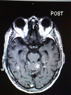


This is an exam I did earlier in the semester that I found interesting. The patient was an 85 yr old man with history of lung cancer. The patient had a screening MRI of the brain about six months prior to the one we did in October to observe for mets due to mental status changes. His GFR was low and the radiologists had the tech do the study without contrast when usually anyone with a cancer history has studies with unless their are contradictions like kidney function. This time the patients GFR was still low but not as low as it was and this radiologist gave us the go ahead to inject contrast due to the patients history. These images are not the best because I took them off the screen at work with my camera phone but you can still see what I am talking about. In the pre images you can see maybe one are that might be something but might be atrophy due to age, it is uncertain if it is a pathology or metastatic disease. Then the injection is given and all I could say was oh my gosh!! All I could keep comparing this man's brain to was a dalmation dogs spots. It was unreal what you could see with the contrast enhancement that you couldn't without it! I wondered if all the mets had been there six months prior when contrast was not used? Look at the images and you will see why I was so shocked at the differences. They didn't upload in the order I wanted them to but the bottom two are the pre images.
.jpg)
No comments:
Post a Comment